Conservative Hallux Valgus Treatment
Overview
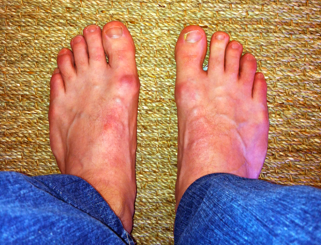 A bunion, also known as hallux valgus, results when the big toe points towards the second toe and results in inflammation of the tissue surrounding the joint. The inflammation causes the joint to become swollen and tender, making everyday activities like walking or jogging very painful. Bunions are much more common in females than in men. This leads most to believe that bunions are often time caused by wearing tight fitting shoes or heels. Genetics also plays an important role in determining the individual?s susceptibility to developing the condition.
Causes
Bunions result from the long bone in the foot (metatarsal) and the big-toe bone becoming misaligned. The causes are likely to be a combination of genetics, wearing ill-fitting shoes, and the way that we walk or run. Arthritis sufferers are also prone to bunions.
Symptoms
If you have a bunion, you may have pain or stiffness of your big toe joint, swelling of your big toe joint, difficulty walking, difficulty finding shoes that fit. These symptoms may be caused by conditions other than bunions, but if you have any of these symptoms, see your doctor.
Diagnosis
Looking at the problem area on the foot is the best way to discover a bunion. If it has the shape characteristic of a bunion, this is the first hint of a problem. The doctor may also look at the shape of your leg, ankle, and foot while you are standing, and check the range of motion of your toe and joints by asking you to move your toes in different directions A closer examination with weight-bearing X-rays helps your doctor examine the actual bone structure at the joint and see how severe the problem is. A doctor may ask about the types of shoes you wear, sports or activities (e.g., ballet) you participate in, and whether or not you have had a recent injury. This information will help determine your treatment.
Non Surgical Treatment
Before treatment of a painful bunion can begin, medical evaluation is needed. There are a number of other causes of pain in the big toe such as osteoarthritis, rheumatoid arthritis, infection and gout. Circulatory problems not only cause pain, but may also cause serious complications if surgery is attempted. Diabetes and cigarette smoking may diminish healing potential and increase the risk of infection. 
Surgical Treatment
An operation may be advised if a change of footwear does not ease symptoms. The aim of the operation is to straighten the joint as much as possible and relieve pain. It is not usually done just to improve appearance. It can be done using a local or a general anaesthetic and you are usually out of hospital the same day. The operation chosen by the specialist depends on the severity of the bunion, the shape of your foot, and other factors, such as if you have arthritis in the joint. There are many different types of operation which are used to treat bunions. These range from operations to trim parts of the joint, to a total artificial replacement of the big toe joint (similar to a knee or a hip replacement). A common surgical procedure used is called a scarf osteotomy (osteotomy means a cut in the bone). An operation is usually successful at easing symptoms, but not in all cases. It is not always possible to relieve the pain completely or make the toe perfectly straight. Your specialist will be able to advise on the pros and cons of surgery, and the success rate of the chosen operation. Continued pain, infection and the bunion returning are some of the complications of surgery of which you need to be aware. You will need to continue to wear sensible, wide-fitting shoes for six months after the operation.
Over-Pronation
Overview
Overpronation and underpronation describe general foot movements. These terms do not necessarily describe a medical problem with a foot. For example, you can overpronate and not have any problems or symptoms at all. It is important to have your foot structure and symptoms adequately assessed by your prescribing physician and a qualified practitioner. Once the underlying conditions and mechanical faults are assessed, an appropriate treatment plan including possible orthotic and footwear recommendations can be made.
Causes
There are many biomechanical issues that can contribute to excessive pronation, including weak foot intrinsic muscles, limited ankle dorsiflexion mobility and calf flexibility, weak ankle invertor muscles (e.g. posterior tibialis), weak forefoot evertor muscles (peroneus longus), poor hip strength and control, Anterior pelvic tilting, heel InversionIn a person who overpronates, the heel bone goes into an everted position meaning that it turns out away from the midline of the body. The opposite motion of eversion is inversion. Inversion is a motion that needs to be controlled to prevent the foot from excessively pronating.
Symptoms
It is important to note that pronation is not wrong or bad for you. In fact, our feet need to pronate and supinate to achieve proper gait. Pronation (rolling inwards) absorbs shock and supination (rolling outwards) propels our feet forward. It is our body?s natural shock-absorbing mechanism. The problem is over-pronation i.e. the pronation movement goes too deep and lasts for too long, which hinders the foot from recovering and supinating. With every step, excess pronation impedes your natural walking pattern, causing an imbalance in the body and consequent excessive wear and tear in joints, muscles and ligaments. Some common complaints associated with over-pronation include Heel Pain (Plantar Fasciitis) ,Ball of foot pain, Achilles Tendonitis, Shin splints, Knee Pain, Lower Back Pain.
Diagnosis
Do the wet foot test. Get your feet wet and walk along a paved surface or sand and look at the footprints you leave. If you have neutral feet you will see a print of the heel with a thin strip connecting to your forefoot, but if you're overpronating your foot print will look a bit like a giant blob with toes.
Non Surgical Treatment
Overpronation is usually corrected with orthotics and/or strengthening exercises for the tibialis posterior. Massage treatment can relieve myofascial trigger points in the tibialis posterior, and other muscles, and address any resulting neuromuscular dysfunction in the leg or foot. Biomechanical correction of overpronation might require orthotics, neuromuscular reeducation, or gait retraining methods, as well. Stretching the gastrocnemius and soleus muscles will reduce hypertonicity in these muscles and also is essential for effective treatment. Because of impacts throughout the remainder of the body, the detrimental effects of overpronation should not be overlooked.
Surgical Treatment
Depending on the severity of your condition, your surgeon may recommend one or more treatment options. Ultimately, however, it's YOUR decision as to which makes the most sense to you. There are many resources available online and elsewhere for you to research the various options and make an informed decision.
Can One Treat Calcaneal Apophysitis In The Home?
Overview
Severs? disease usually presents with pain in either one or both of a sufferer?s heels. The area can be sore or tender, particularly first thing in the morning or after squeezing. Because the pain is focussed on the heel, an important part of the foot that makes contact with the ground through virtually all movement, sufferers often have to limp to alleviate their discomfort. The pain of Severs? disease is at its worst after any exertion that involves contact between a heel and the ground, particularly strenuous exercise like running or sport. The condition is caused by the wear and tear of structures in the heel, most significantly the heel bone and any attached tendons. Severs? disease is prevalent in young children who are extremely active, particularly as the heel and its attached tendons are still growing in the age group the condition most commonly affects (7-14).
Causes
Growth plates, also called epiphyseal plates, occur at the end of long bones in children who are still growing. These plates are at either end of growing bones, and are the place where cartilage turns into bone. As children grow, these plates eventually become bone (a process called ossification). During a growth spurt, the bone in the heel may outpace the growth of the muscles and tendons that are attached to the heel, such as the Achilles tendon. During weight bearing, the muscles and tendons begin to tighten, which in turn puts stress on the growth plate in the heel. The heel is not very flexible, and the constant pressure on it begins to cause the symptoms of Sever?s disease. Sever?s disease is common, and it does not predispose a child to develop any other diseases or conditions in the leg, foot, or heel. It typically resolves on its own.
Symptoms
Pain is usually felt at the back of the heel and around the sides of the heel. If you squeeze the back of the heel from both sides simultaneously and pain is experienced Sever?s disease is more than likely present.
Diagnosis
A doctor or other health professional such as a physiotherapist can diagnose Sever?s disease by asking the young person to describe their symptoms and by conducting a physical examination. In some instances, an x-ray may be necessary to rule out other causes of heel pain, such as heel fractures. Sever?s disease does not show on an x-ray because the damage is in the cartilage.
Non Surgical Treatment
Your child may need to change or stop doing the activities that cause pain until the growth plate heals. Your child needs to wear shoes with good padding in the heels and good arch support. Special shoes or shoe inserts may help. Pain from Sever's disease may last weeks to months. The pain may come back if your child returns to sports or strenuous activities too soon.
Prevention
Properly stretching to maintain flexibility is effective for preventing Sever's disease. Stretches should target the calves, heel cords, and hamstrings. Your child should do the appropriate stretches two or three times a day, holding the stretch for about 20 seconds each time. Ask your child's doctor for specific exercise instructions. Generally, doctors advise stretching both legs, even if the pain is confined to one heel. It's also helpful to strengthen the shin muscles by having your youngster pull his toes in with a rubber exercise band or a piece of tubing and then stretch them forward. Assist your child in doing 15 repetitions of this exercise, three times a day. Having your child wear shoes with good shock absorbers and avoid running on hard surfaces as much as possible should also help prevent the condition.
Achilles Tendon Rupture Repair And Physical Therapy
Overview
 An Achilles tendon rupture is also known as a rupture of the gastrocnemius tendon, or the common calcanean tendon. The tendon is actually composed of 5 different tendons, the two most important being the superficial digital flexor and gastrocnemius tendons. The gastrocnemius tendon is the largest of these, and is the most powerful extensor of the hock (ankle) joint. Both the superficial digital flexor and gastrocnemius tendons attach to the heel bone, called the calcaneus bone. A rupture of the Achilles tendon may be a partial tear, which means just the gastrocnemius is torn, or a complete tear, in which all five tendons have been torn. (show diagrams, normal anatomy vs partial vs complete tears).
Causes
Factors that may increase your risk of Achilles tendon rupture include some of the following. Age. The peak age for Achilles tendon rupture is 30 to 40. Sex. Achilles tendon rupture is up to five times more likely to occur in men than in women. Recreational sports. Achilles tendon injuries occur more often during sports that involve running, jumping, and sudden starts and stops, such as soccer, basketball and tennis. Steroid injections. Doctors sometimes inject steroids into an ankle joint to reduce pain and inflammation. However, this medication can weaken nearby tendons and has been associated with Achilles tendon ruptures. Certain antibiotics. Fluoroquinolone antibiotics, such as ciprofloxacin (Cipro) or levofloxacin (Levaquin), increase the risk of Achilles tendon rupture.
Symptoms
Typically patients present with sudden onset of pain and swelling in the achilles region, often accompanied by a audible snap during forceful dorsiflexion of the foot. A classic example is that of an unfit 'weekend warrior' playing squash. If complete a defect may be felt and the patient will have only minimal plantar flexion against resistance.
Diagnosis
A physician usually can make this diagnosis with a good physical examination and history. X-rays usually are not taken. A simple test of squeezing the calf muscles while lying on your stomach should indicate if the tendon is still connected (the foot should point). This test isolates the connection between the calf muscle and tendon and eliminates other tendons that may still allow weak movement. A word of caution, Achilles tendon rupture is often misdiagnosed as a strain or minor tendon injury. Swelling and the continuing ability to weakly point your toes can confuse the diagnosis. Ultrasound and MRI are tests that can assist in difficult diagnosis. Depending on the degree of injury, these tests can also assist in determining which treatment may be best.
Non Surgical Treatment
Initial treatment for sprains and strains should occur as soon as possible. Remember RICE! Rest the injured part. Pain is the body's signal to not move an injury. Ice the injury. This will limit the swelling and help with the spasm. Compress the injured area. This again, limits the swelling. Be careful not to apply a wrap so tightly that it might act as a tourniquet and cut off the blood supply. Elevate the injured part. This lets gravity help reduce the swelling by allowing fluid and blood to drain downhill to the heart. Over-the-counter pain medication is an option. Acetaminophen (Tylenol) is helpful for pain, but ibuprofen (Motrin, Advil, Nuprin) might be better, because these medications relieve both pain and inflammation. Remember to follow the guidelines on the bottle for appropriate amounts of medicine, especially for children and teens. 
Surgical Treatment
Surgery offers important potential benefits. Besides decreasing the likelihood of re-rupturing the Achilles tendon, surgery often increases the patient?s push-off strength and improves muscle function and movement of the ankle. Various surgical techniques are available to repair the rupture. The surgeon will select the procedure best suited to the patient. Following surgery, the foot and ankle are initially immobilized in a cast or walking boot. The surgeon will determine when the patient can begin weightbearing. Complications such as incision-healing difficulties, re-rupture of the tendon, or nerve pain can arise after surgery. Whether an Achilles tendon rupture is treated surgically or non-surgically, physical therapy is an important component of the healing process. Physical therapy involves exercises that strengthen the muscles and improve the range of motion of the foot and ankle.
Prevention
There are things you can do to help prevent an Achilles tendon injury. You should try the following. Cut down on uphill running. Wear shoes with good support that fit well. Always increase the intensity of your physical activity slowly. Stop exercising if you feel pain or tightness in the back of your calf or heel.
What Is The Cause Of Adult Aquired FlatFeet
Overview
Adult acquired flatfoot deformity or posterior tibial tendon dysfunction is a gradual but progressive loss of ones arch. The posterior tibial muscle is a deep muscle in the back of the calf. It has a long tendon that extends from above the ankle and attaches into several sites around the arch of the foot. The muscle acts like a stirrup on the inside of the foot to help support the arch. The posterior tibial muscle stabilizes the arch and creates a rigid platform for walking and running. If the posterior tibial tendon becomes damaged or tears the arch loses its stability and as a result, collapses causing a flatfoot. Adult flatfoot deformity can occur in people of all ages and gender however, it occurs most commonly in sedentary middle aged to elderly females. There are several risk factors for posterior tibial tendon dysfunction that include: obesity, steroid use, systemic inflammatory diseases such as rheumatoid arthritis, trauma, being born with a low arch, and diabetes. It occurs most commonly in one foot however, it can occur in both feet especially in people with systemic diseases such as diabetes and rheumatoid arthritis. 
Causes
Adult acquired flatfoot is caused by inflammation and progressive weakening of the major tendon that it is responsible for supporting the arch of the foot. This condition will commonly be accompanied by swelling and pain on the inner portion of the foot and ankle. Adult acquired flatfoot is more common in women and overweight individuals. It can also be seen after an injury to the foot and ankle. If left untreated the problem may result in a vicious cycle, as the foot becomes flatter the tendon supporting the arch structure becomes weaker and more and more stretched out. As the tendon becomes weaker, the foot structure becomes progressively flatter. Early detection and treatment is key, as this condition can lead to chronic swelling and pain.
Symptoms
The symptoms of PTTD may include pain, swelling, a flattening of the arch, and an inward rolling of the ankle. As the condition progresses, the symptoms will change. For example, when PTTD initially develops, there is pain on the inside of the foot and ankle (along the course of the tendon). In addition, the area may be red, warm, and swollen. Later, as the arch begins to flatten, there may still be pain on the inside of the foot and ankle. But at this point, the foot and toes begin to turn outward and the ankle rolls inward. As PTTD becomes more advanced, the arch flattens even more and the pain often shifts to the outside of the foot, below the ankle. The tendon has deteriorated considerably and arthritis often develops in the foot. In more severe cases, arthritis may also develop in the ankle.
Diagnosis
The diagnosis of tibialis posterior dysfunction is essentially clinical. However, plain radiographs of the foot and ankle are useful for assessing the degree of deformity and to confirm the presence or absence of degenerative changes in the subtalar and ankle articulations. The radiographs are also useful to exclude other causes of an acquired flatfoot deformity. The most useful radiographs are bilateral anteroposterior and lateral radiographs of the foot and a mortise (true anteroposterior) view of the ankle. All radiographs should be done with the patient standing. In most cases we see no role for magnetic resonance imaging or ultrasonography, as the diagnosis can be made clinically.
Non surgical Treatment
The following is a summary of conservative treatments for acquired flatfoot. Stage 1, NSAIDs and short-leg walking cast or walker boot for 6-8 weeks; full-length semirigid custom molded orthosis, physical therapy. Stage 2, UCBL orthosis or short articulated ankle orthosis. Stage 3, Molded AFO, double-upright brace, or patellar tendon-bearing brace. Stage 4, Molded AFO, double-upright brace, or patellar tendon-bearing brace. 
Surgical Treatment
Surgical intervention for adult acquired flatfoot is appropriate when there is pain and swelling, and the patient notices that one foot looks different than the other because the arch is collapsing. As many as three in four adults with flat feet eventually need surgery, and it?s better to have the joint preservation procedure done before your arch totally collapses. In most cases, early and appropriate surgical treatment is successful in stabilizing the condition.